

Biology is Too Hard
And the Mark Schemes are never clear
Muscle Structure & Contraction
Remember muscle tissue is not made up out of individual cells. This is because if it were there would be lines of weakness. Instead all of the cells fuse together and share their cytoplasm and nuclei - this is known as sarcoplasm.
Muscle tissue has a large concentration of mitochondria and rough endoplasmin reticulum.
Each muscle fiber is made up out of things called myofibrils. Each myofibril contains 2 proteins - actin and myosin.


The picture to the right shows how the actin and myosin filaments are arranged in a myofibril. For your exam you have to know
1) Where the I Bands and A bands are
2) Where the H Zone is
3) Where the Z lines are
The myofibril beneath shows the M line, which is in the centre of the A band.

This is a cross section through part of a muscle fiber. It shows how actin and myosin filaments are arranged in a myofibril. The thick ones are myosin protein filaments and the thin ones are actin protein filaments.


Above and to the left are sarcomeres. These are the basic units of muscles. It is here where we see the actin filaments sliding past the myosin filaments. The above picture is taken with an electron microscope and shows you the light and dark banding. You can clearly make out the Z lines (sometimes called Z disks). You can also see light and dark regions. The light regions are where there is just one type of filament and the dark regions are where the actin and myosin filaments overlap. The M line is actually a protein called myomesin that holds the myosin filaments together and keeps the myosin filaments in place.

The Sliding Filament Theory
The two protein filaments to the left are actin and myosin. Myosin in the red and actin in the blue. On the actin you can see the green tropomyosin protein wound around the actin fibre and the yellow dots on them (think they are yellow) must be Troponin.

1.) Once the action potential arrives at a neuromuscular junction it depolarises the membrane. This wave of depolarisation is carried deep into the myofibrils through T - tubules. (T stands for transverse). The picture to the right shows you the T-tubules in yellow.
2.) The tubules are in contact with the sarcoplasmic reticulum - the depolarisation causes Ca2+ ions (I cant find out how to get the 2+ above the Ca) to be released from the sarcoplasmic reticulum. The sarcoplasmic reticulum actively uptakes Ca2+ from the cytoplasm (or sarcoplasm in muscle).

3.) The Calcium ions bind to Troponin, which causes the tropomyosin filament to move revealing the myosin binding sites.

4.) The next thing to happen is shown in step 1 of the opposite diagram. The myosin head is free to bind with the actin filament at the exposed binding site. This forms a cross bridge. Remember ATP & Pi are still attached to the head.
5.) Pi is released first causing the myosin head to pivot and bend - this is the power stroke. It slides the actin filament towards the M line. Then ADP is released.
6.) ATP is now needed to break the cross-bridge. It does this by binding to the myosin head. To 're-cock' the myosin head Ca2+ ions activate the enzyme ATPase, which hydrolyses ATP - ADP & Pi - remember these stay bound to the myosin head. This provides the energy for the 're-cocking'.
7.)So long as there is nervous stimulation then the myosin head will attach to an actin filament further down and so the cycle is repeated.
Types of Muscle
There are 2 muscle types that you need to know
1) Slow - Twitch Fibres
2) Fast - Twitch Fibres
Slow Twitch
These contract more slowly and are less powerful. They are adapted for endurance work. Therefore they try to keep in aerobic respiration for as long as possible. They have
-
A large store of myoglobin (a protein similar to haemoglobin - it lives in muscle cells and stores oxygen)
-
A large supply of glycogen
-
Rich supply of blood vessels
-
Loads of Mitochondria
Fast Twitch
These muscles contract rapidly and produce powerful contractions. They are adapted by having
-
thicker and more numerous myosin filaments
-
Higher concentration of enzymes involved in anaerobic respiration
-
A large store of Phosphocreatine
Muscle contraction requires ATP for
1) 're-cocking' the myosin head
2) Actively re uptaking Ca2+ ions into the sarcoplasmic reticulum.
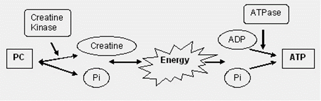
Active muscle has a large demand for ATP then. When ATP can't be regenerated from ADP and Pi in oxidative phosphorylation - because all the oxygen has been used up - the muscles still need some way of making ATP anaerobically. Muscle tissue stores phosphocreatine and it is used in the diagram above. The cleaving of PC provides the energy to combine the Pi with ADP.
This is a great animation for showing you muscle contraction.